


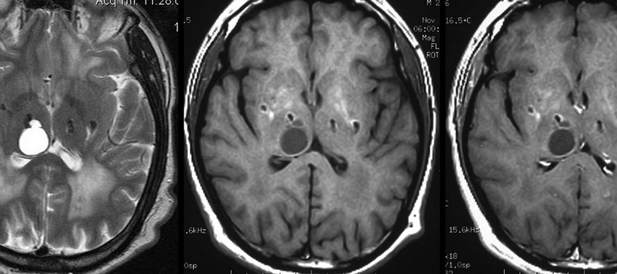
What is a Brain Tomography?
Brain Tomography; There are devices that are changing and developing every day in the field of health. One of these devices is a device known as brain tomography. Brain tomography is used for a sensitive organ such as the brain.
How to use brain tomography, which is often used by the department of radiology, in a real sense. The root of the word tomography comes from the Greek.
It is a combination of the Greek word for slice or cut and the words save, write. The procedures performed by radiology specialists are divided into two main groups. One of them is referred to as computed tomography, while the other is referred to as normal tomography.
In a tomography requested by your doctor, a radiology specialist or radiology technician performs a tomography scan procedure. Here, the process takes place for the desired region. A detailed conclusion is prepared after the film is shot. The prepared result is sent to the physician at a later stage.
Tomography, which is used as a diagnostic tool, is performed using x-ray rays. The process takes place for the distressed area on the body scanned by these rays. During the shooting, the bones, vessels and organs in the body are checked in sections.
Unlike X-ray, computer tomography or normal tomography is evaluated in detail. after the image presented in 3-Dimensional sections, the physicians inform how the treatment to be applied will take place.
How is a Brain Tomography performed?
After the necessary examination, the patient who is requested tomography is directed to the area where the procedure will be performed. It is necessary not to eat for about 6 hours before the procedure to be performed. In the same way, for the brain tomography procedure, it is necessary not to drink water 4 hours before the procedure.
The patient who comes to the department takes off all the clothes he has on. In the same way, he also removes all the jewelry that is on the patient. At this time, all items such as hairpins or dentures or glasses and jewelry located in the hair must be removed.
In this section, the patient already receives the necessary warnings by the responsible physician. Then the preparations are completed and the patient is laid on a stretcher.
Gym in meyreuil, municipal sports complex – (bouches-du-rhone) buy tamoxifen 20 tamoxifen citrate nolvadex with uk shipping sapeurs pompiers barracks (weight room / cardiotraining room), fournès (30210) – gard.
Patients who want to have a brain tomography are usually located at a fixed point, that is, the tomography tool should move while the patient is standing still. In some different models, the device stands still and the stretcher rotates. At the same time, in order for the patient to be comfortable and for the image to be taken to be healthy, the patient is fixed with pillows and connecting straps.
During the time of the shooting, the patient is left alone with the device that will perform a tomography scan. At this time, no one else is with the patient. However, the specialist who will take the patient’s shot will be in a position to monitor the patient throughout the shooting.
The image to be obtained must be clear. It is quite important that this image is clear and not repeated. Reshoots will be made in cases where the image is not clear.
What is a Medicated Brain Tomography?
In addition to standard tomography, medicated tomography is performed to obtain clearer images. The patient’s condition is also important for the shooting, which will be performed by administering medication to the targeted area. Medication is given according to the patient. After this medication is given, a brain tomography scan is performed.
The required drug efficiency for the areas where brain tomography is located varies depending on the patient’s condition. During the shooting, the drugs act in the body. In this way, according to the action of drugs, the disease will be diagnosed much better. Thus, the desired treatment will be started in a short time.
After the necessary studies, the radiology specialist prepares a report for the patient. The prepared report is presented to the physician. The patient is diagnosed as a result of brain tomography controlled by the physician.
After the diagnosis is made, the necessary treatment is performed for the patient. Radiology specialists do not make any comments for diagnosis and treatment, and specialists do this work.
Brain Tomography Prices 2022
You can contact us immediately to get information about brain tomography prices 2022.
Yorumlar (Henüz yorum yapılmamış)
Henüz yorum yapılmamış