


In cases requiring cephalometric, orthodontic diseases or orthognathic surgical intervention, clinical observation may not always be sufficient. For this reason, X-ray machines have been started to be used to take an X-ray of the facial area and allow the doctor to have a detailed idea about the disease.
In this way, the head is viewed from different angles and it is possible to take X-rays of tissues and bones. These machines, which detect the location and extent of the problem, have a wide range of uses. This has led to the fact that it has become a method that is often used by doctors to diagnose diseases.
This type of X-ray is used to determine the cause of pain in the jaw and teeth, the cause of which cannot be determined by clinical observations, or to view the entire region during operations such as dental caries, root canal treatment. In this way, it is possible to detect diseased structures such as cysts, tumors or bruises that the doctor cannot see.
This increases the chances of treatment applied by the doctor. Obtaining the results in a short time is a very practical solution for diseases that require urgent intervention. By imaging all of the tissues, the cystic structures that will form in the jaw and roots are determined in advance and the chance of treatment is obtained.
How is a Cephalometric X-Ray Taken?
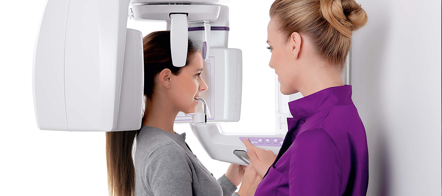
For X-ray taking, the sensors should be positioned parallel to the patient’s midsagittal plane. For this reason, the patient puts his chin in the place indicated by the doctor on the device. In order for the sensors to detect the film properly during shooting, the patient must not move.
Sensors located around the patient, who keeps his head still, create a radiograph of the head by sending X-Ray rays. There are filters that absorb some of these rays during shooting. The reason for this is to ensure that the tissues are also visible on the radiograph.
In films taken with cephalometric X-rays, a lower dose of radiation is used compared to other types of X-rays. This significantly reduces the amount of radiation received into the body, and the head, which is a sensitive area, is less exposed to X-Ray rays.
This machine, which shoots in size equal to the size of the head, provides an opportunity to assess the size of the bones, their position relative to each other, their dimensional differences. By taking the head from both the front profile and the side profile, it allows all the depths in the head to be displayed.
The filming takes only a few seconds and then the results come out immediately. This type of X-ray, which does not require any preparation, does not affect the patient after the shooting either. The results can be shared with the patient as a CD or online according to the patient’s request.
How is Cephalometric Evaluation Performed?
Cephalometric assessment is performed in two ways. When filming, the facial area is taken both from the front and from the side. The reason for this is that the machine captures images in two dimensions and is not able to capture surfaces of different depths from one angle. In the filmed film, the position of the bones relative to each other is evaluated first.
When any abnormality is detected, the source of the problem is investigated and the necessary treatment method is applied. Secondly, soft tissue and hard tissue assessment is performed. Separate evaluation of two tissue types gives the same diagnostic results.
Not only the film results should be included in the evaluation, but also the clinical observations. Based on the amount of radiation received by patients undergoing filming, filming should not be performed as an extra in evaluations where clinical observation is sufficient.
However, if necessary, the doctor may request a film next to the clinical observation and make the evaluation accordingly. Sometimes discrepancies arise between two observations. Decoupling. This does not always mean that there is an abnormality, sometimes an excessively thick or thin soft tissue located in the upper layer can also cause this discrepancy.
For this reason, clinical observations should be taken into account instead of film results when preparing a treatment plan. The margin of error is higher for treatments performed only with film results.
Cephalometric X-Ray Prices 2022
You can contact us immediately to get information about cephalometric X-ray prices 2022.
Yorumlar (Henüz yorum yapılmamış)
Henüz yorum yapılmamış